QUICK APPOINTMENT FORM

What is Glaucoma (Eye Pressure)?
Glaucoma is a serious disease that can cause loss of vision due to damage to the visual nerve as a result of chronically high intraocular pressure. The most common type of glaucoma is open-angle glaucoma, which has a long and insidious course. Open-angle glaucoma accounts for 85 to 90 per cent of all glaucoma patients. The patient’s eye pressure suddenly rises to very high levels, causing severe eye pain, headache, vomiting, nausea and excessive red eye. This is an emergency and very severe condition. It is easy to recognise by ophthalmologists.
Response to intensive treatment is immediate. Treatment is surgical (conventional or laser). In addition, patients at risk of angle closure can be easily identified in advance and precautions can be taken. There is also a rare type called secondary glaucoma. Here, there is an increase in blood pressure in the eye as a result of another eye disease (cataract, trauma, intraocular inflammation, intraocular haemorrhage). The treatment is the treatment of blood pressure together with the disease.
In the human eye, a fluid that nourishes the intraocular tissues is continuously produced and leaves the eye through the canals in the eye and passes into the blood circulation. If there is stenosis, adhesion or blockage in these canals leaving the eye, the outflow of the fluid slows down and the pressure inside the eye increases.
In Which Range Should Eye Pressure(Glaucoma) Be?
This increases the pressure (blood pressure) of the eye. Normally, eye pressure is between 12-21 mm Hg. When the blood pressure is between 25-40 mm Hg, it is usually asymptomatic, so it is caught by chance during an eye examination. Sometimes there may be complaints such as pain and blurred vision. It is usually seen over the age of 40, but it can even occur in infants. 2-3 of every 100 people over the age of 40 have eye pressure.
Blood pressure deteriorates the eye nerves and tissues over time, reduces vision and may lead to blindness. It has nothing to do with food. It is hereditarily common in some families. Those with a family history of glaucoma should be more careful. It is more common in high myopia (above 5 degrees) and high hyperopia (3 degrees and above). For the diagnosis, blood pressure is measured several times and examined. Investigations such as visual field, optic nerve topography are performed.
Those with borderline eye pressure (20-23) should be checked more frequently. If eye pressure is not treated, the patient cannot see his/her surroundings even if he/she can see where he/she is looking. He/she may not be able to recognise people or cars approaching from his/her right or left. In the treatment, drops are applied once or several times a day. Sometimes laser treatment is performed. If these do not reduce blood pressure and prevent damage, surgery is performed. Since glaucoma does not give many symptoms and can lead to blindness, it is important to go for regular eye examinations.
What are the Causes and Symptoms of Glaucoma?
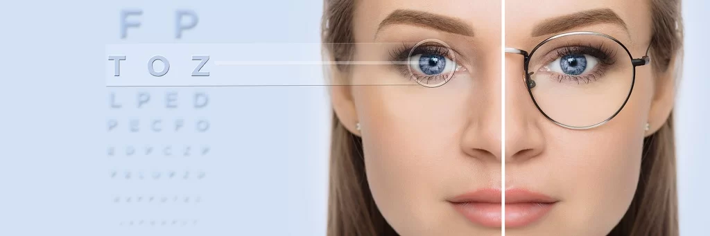
Open-angle glaucoma, which is insidious or does not give any symptoms if it is not advanced, is usually diagnosed by measuring eye pressure as part of a routine examination in patients presenting with near vision complaints. High eye pressure is a suspicious finding in diagnosis and measurements; however, it cannot be decided only by looking at it. Because there are patients who have visual nerve damage even though their eye pressure is low, and there are also patients who do not show nerve damage even though their blood pressure is measured high.
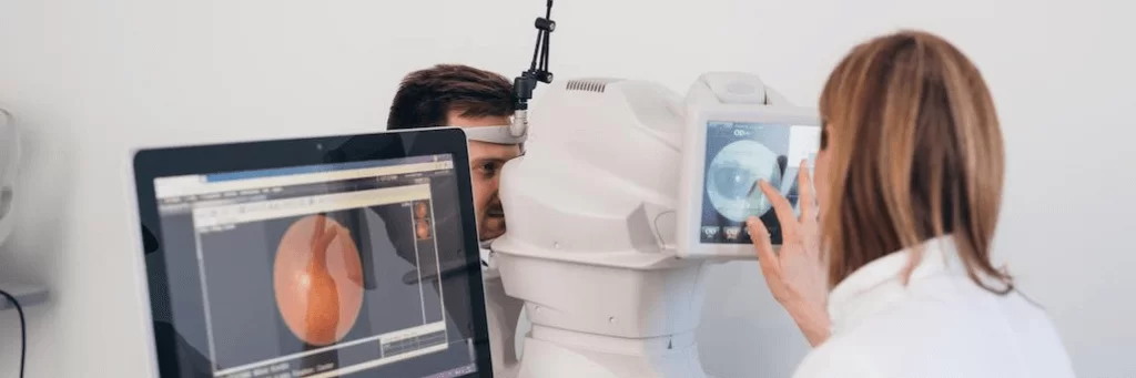
Visual damage can be detected by various methods. In the examination, the optic nerve can be examined with devices such as direct biomicroscope and signs of damage can be detected. In suspicious cases, fundus photographs can be taken. In addition, OCT (Optical Coherence Tomography) and NFA (Neuro-Fuzzy Algorithmic) methods can detect the damage of the optic nerve and surrounding nerve fibres at microscopic level. Visual field examination is a must. This is a functional and very valuable test that shows us the areas with visual field loss in the eye like a map. 90 per cent of patients are over the age of 40. The risk of glaucoma is 10 times higher in patients with a genetic predisposition.
Therefore, eye pressure should be measured at any age during normal examination. In order to recognise this disease and to diagnose it early, an annual eye examination is recommended after the age of 40. If the disease has an insidious course and is left to its own devices, i.e. if it is not diagnosed and never treated, it causes visual damage up to the complete loss of even light in 10-12 years. Sometimes it even leads to surgical removal of the eye.
 1")
Is Glaucoma Treatable?
Glaucoma is a disease that can be treated. Especially if it is diagnosed early, this is even easier. When the diagnosis is made late, the eye is destroyed due to years of high blood pressure and its endurance decreases. Even if the blood pressure decreases, visual field losses continue and sometimes it is not possible to save the eye even with maximum treatment. Therefore, it is very important to comply with the treatment after early diagnosis in this disease, that is, to use the drops regularly and to come to the controls.
Glaucoma (Eye Pressure) Treatment
Glaucoma can be treated in three different ways;
- Medication
- Laser
- Operation
Drug treatment: There are many drops that act by various mechanisms in the treatment of glaucoma. Intraocular pressure is tried to be reduced with one drug. If it is not enough, a second drop is added. If the eye pressure does not decrease again, the third drop is added or other treatment methods are used.
Laser Treatment: Laser treatment is a last treatment option that can be applied before surgery in patients who do not respond adequately to drug treatment. Laser treatment can reduce intraocular pressures that are not too high to normal levels and can safely reduce medications in patients who are at risk of taking medication. The duration of effect is usually 2-3 years. Then intraocular pressure may rise again.
Operation: If the patient with glaucoma has tried all possible methods, but the pressure cannot be reduced or visual field loss persists, surgery is mandatory. Glaucoma surgery is performed under local anaesthesia unless the patient is a child. Regular eye examination once a year is necessary for early diagnosis.
 2")
Minimally Invasive Glaucoma Surgery in Glaucoma (Eye Pressure) Treatment
The goal of minimally invasive glaucoma surgery, a new option in the treatment of glaucoma, is to prevent or reduce ocular nerve damage due to glaucoma. Today, the most common surgery for the treatment of glaucoma is trabeculectomy. This surgery is considered a major surgical procedure for the eye. Although this surgery effectively reduces intraocular pressure, there may be some unwanted complications after surgery.
In recent years, a minimally invasive glaucoma surgery technique has been developed. With this technique, intraocular pressure is effectively reduced and postoperative complications are considerably reduced.
In this technique, which is performed under local anaesthesia, no stitches are placed in the eye. The procedure takes 20-30 minutes. The eye remains closed with a bandage for one day. After one day, the patient can return to daily life. Healing drops should be used for one month. After the operation, 70% of the patients can get rid of the drops used in the treatment of glaucoma. In 30% of patients, the number of drops used is reduced. This technique can be applied alone or in combination with cataract surgery.
Which patients do we recommend this surgery?
- Patients whose intraocular pressure does not decrease to a safe level despite maximum medication.
- Patients who develop side effects (allergies, etc.) due to the drops they use.
- Patients who cannot apply glaucoma treatment drops.
 3")
How Much Is Glaucoma Treatment Price?
Glaucoma (Eye Pressure) treatment prices vary according to the underlying cause, special examination and detailed measurements to be made to your eye depending on the operation to be performed. The treatment plan to be created by our specialist ophthalmologist depending on the detailed examination results may vary from person to person.
The above information is for informational purposes. If you have any medical concerns or questions, please make an appointment with our ophthalmologists.